Tag Archives: parietal evolution

A full view of the angular gyrus

image (10)

The journal Brain Structure and Function has recently published a collection of articles, a special issue, dedicated to the angular gyrus, a fundamental element of the parietal lobe. The Special Issue: Angular Gyrus is guest edited by Kathleen Rockland, research professor in anatomy and neurobiology, as well as William Graves, associate professor of psychology. The angular gyrus (AG) is an interesting area of association cortex because of its diverse structural and functional connectivity, involved in mathematical, spatial and social cognition, among others. Consequently, many approaches have been employed to study its anatomy and functions, although there is still much to be learned about this cortical region and its evolution. The articles featured in the special issue were published over the course of several months, starting in April. They include both original research articles and reviews, and cover a range of different topics including comparative anatomy, connectivity, cytoarchitecture and cognition. All in all, this special issue is meant to be more than the sum of its parts: informing not only about the structure and function of the angular gyrus, but also about brain organization as a whole.

Tim Schuurman


Neanderthal and modern human parietal lobes

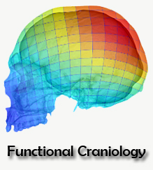
Neanderthals and modern humans have similar cranial capacities but different brain shapes. While our species is characterized by globular braincase and vertically bulging parietal regions, Neanderthals typically have elongated endocrania and flatter parietal regions. Human endocasts usually display smooth surfaces, lacking clear imprints of cortical features, which makes it difficult to define a particular brain region based on homologous landmarks. In this sense, surface semilandmarks are usually applied, but these cannot capture information on changes associated with specific cerebral subdivisions. Now, in collaboration with Philipp Gunz and Simon Neubauer, we combined the two approaches to compare the parietal lobes of modern humans and Neanderthals. The study incorporated landmarks corresponding to elements and boundaries of the parietal lobe into a dense landmark and semilandmark set representing the whole endocranial surface, to obtain a comprehensive analysis of modern human and Neanderthal anatomical differences in shape and size of the parietal cortex and its subregions. The sample included 52 modern humans, from diverse geographical origins, and 8 Neanderthals, namely Amud 1, Feldhofer 1, La Chapelle-aux-Saints 1, La Ferrassie 1, Guattari 1, Saccopastore 1, Spy 1 and Spy 2. We performed two geometric morphometric analyses, one on the whole endocranial configuration and another one on the parietal lobe region only. We also measured the parietal lobe arc and chord, and its surface area. In general, when compared to Neanderthals, the parietal lobes of modern humans are located in a more superior position and are dorsally rounder, longitudinally more extended, and vertically stretched. Our geometrical model showed that modern humans display expanded areas on the dorsal parietal region, towards the parietal-occipital boundary, and on a lateral region between the supramarginal and angular bosses and the intraparietal sulcus. Areas of the intraparietal sulcus are involved in eye-hand and hand-tool coordination, and those of the superior parietal lobule in visuospatial processing. Nonetheless, Neanderthal and modern human morphological ranges overlap, suggesting that species-specific differences might be only by grade.

Sofia Pedro