Tag Archives: morphological integration

Modularity and community detection

Schuurman_Bruner_2023_GraphicalAbstract

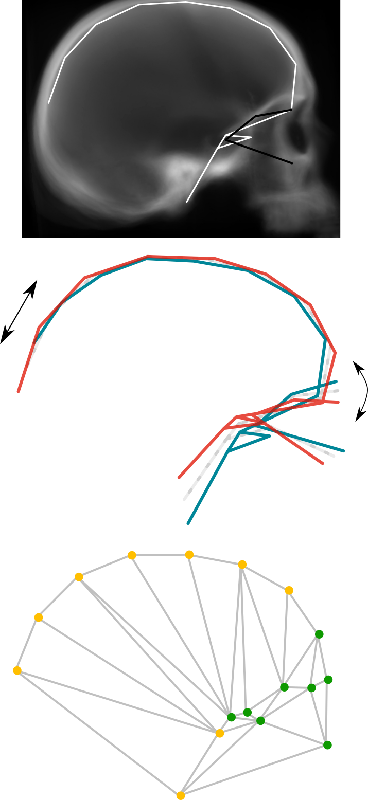
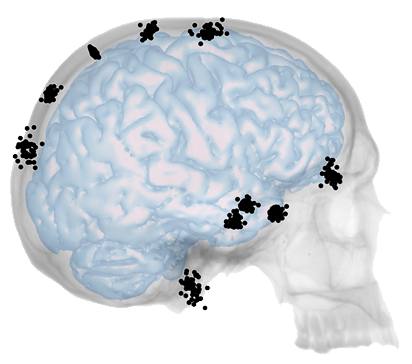
We have recently published a new paper regarding modularity and community detection in the context of human brain anatomical network analysis (Schuurman & Bruner, 2023a). Humans’ morphologically complex brain is spatially constrained by the physical interactions of its elements, and this aspect can be studied using anatomical network analysis. A crucial issue in this framework is modularity, assessed by means of community detection algorithms: the presence of groups of elements undergoing morphological changes in a concerted manner.

Previous works on the subject had produced mixed results: Bruner et al. (2019) and Bruner (2022) found longitudinal (anterior-posterior) modular partitions of the brain, while Schuurman & Bruner (2023b) found a vertical (superior-inferior) modular partition. In the present study, our aim was to test an array of community detection algorithms on a previously designed anatomical network model (Schuurman & Bruner, 2023b), offering a quantitative exploration of modularity to generate well-founded interpretations of the observed morphological organization of the human brain.

The algorithms that were examined comprise the combined method of the spin-glass model and simulated annealing algorithm (SG-SA), a partitional method with an automatic determination of the number of communities; the Louvain method, a heuristic method reliant on Q optimization with no a priori assumptions on community size or number; Infomap, an algorithm that focuses on information diffusion across the network; the generalized topological overlap measure (GTOM), a hierarchical clustering method based on the level of structural equivalence of the network’s nodes; and agglomerative hierarchical clustering based on maximal clique (EAGLE), which identifies initial subsets of completely connected nodes, then merges these subsets hierarchically according to their topological similarity and identifies the ideal partition using Q optimization.

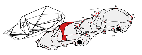
The algorithms that provide the highest quality partitions are SG-SA and the Louvain method. However, all five of them supply useful information. SG-SA, Louvain and Infomap reveal a clear vertical modular partition of the brain: they yield only two superior modules (these span regions as posterior as the middle occipital gyrus) and three inferior modules. Additionally, Informap goes as far as to subdivide the posterior inferior module vertically again, into a superior and an inferior posterior submodule. On the other hand, GTOM and EAGLE, show a more obvious longitudinal division: firstly, they separate the superior modules longitudinally (these only reach as far as the supramarginal gyrus), generating an additional, more posterior, superior submodule; and secondly, many regions incorporated in the inferior modules by SG-SA, the Louvain method and Infomap, are here considered to be part of the superior modules, contributing even further to this sense of anterior versus posterior. In other words, jointly, the community detection algorithms expose the simultaneous occurrence of a longitudinal and a vertical modular partition. This combination mirrors the morphological organization of the enveloping braincase, separated vertically by the distinct developmental processes of the cranial base and vault, and longitudinally by the particular morphogenetic environments of the three endocranial fossae. Overall, results suggest a level of concerted topological reciprocity in the spatial arrangement of craniocerebral anatomical components. Lastly, they posit questions concerning the degree to which structural constraints of the skull and the modular partition of the brain may channel both evolutionary and developmental trajectories.

Tim Schuurman


Spatial packing in the macaque head

jeffery (4)Head morphology reflects, besides function, inherent structural constraints. This is interesting in developmental studies because, as tissues grow and compete for space, structural constraints lead to a modification of the genetically defined adult phenotype. In their latest work, Jeffery and Manson (2023) set out to investigate these geometric alterations in the postnatal stage of head development in the rhesus macaque (Macaca mulatta). Specifically, they used magnetic resonance imaging (MRI) data of 32 male and female individuals aged 0‒3 years old. Their aim was to find the most significant modifier in brain and skull shape out of relative size changes in the brain, eyeballs and masseter muscles, as well as neuronal wiring length, using patterns of covariation.

Results reveal that, in the rhesus macaque, associations of brain and skull shape with eyeball size and neuronal wiring length are weak across the board. However, these findings do not deny the significance of neuronal wiring length as a structural constraint in other species, as the functional threshold for length optimization might not have been reached in the sample in question. Meanwhile, brain and skull shape are shown to be influenced greatly by the relative size of the masseter muscles and, surprisingly, the face and basicranium, rather than the brain. In other words, this research contributes to the idea that skeletal growth should be considered properly in models of spatial packing, instead of focusing almost exclusively on the brain. In conclusion, the trajectory and efficacy of spatial-packing events in the head, as well as the transition from a spatially modular system to a more integrated one, is probably dependent on the combined developmental timings of the brain, masticatory muscles, and cranium.

Tim Schuurman


Human brain network analysis

Schuurman&Bruner_Prensa_CENIEH_Imagen_1

We have published a new study on the evolution of human brain morphology by means of anatomical network analysis (Schuurman & Bruner, 2023), which permits modeling physical interactions among the brain’s morphological elements. These physical interactions inform about geometric constraints which, once untangled, reveal key regions as well as broader phenotypic patterns in the brain’s spatial arrangement. Altogether, this information helps us to better understand the ontogeny and evolution of human brain morphology.

A preliminary study published on this topic (Bruner et al. 2018) highlighted the presence of a parieto-occipital spatial block. Two subsequent models (Bruner et al. 2019 and Bruner 2022) suggested an antero-posterior morphological partition of the brain. The former analysis concerned the brain’s macroscopical cortical regions, while the latter was based on the Brodmann’s map. This new work pushes this approach further by including 101 cortical and subcortical macroanatomical brain regions across both hemispheres and over 300 interactions.

Results reveal that the parahippocampal gyrus is the region with the most physical interactions, a measure of local morphological integration, and the highest betweenness centrality, a measure of global integration. Moreover, this region also holds the most central position within the network. This is likely to be due to its dimensions and elongated shape. Other relevant regions include the anterior lobe of the cerebellum and the ventral portion of the midbrain. Due to their topological properties, all three regions are subjected to geometrical constraints that are likely to limit their evolutionary rate, at least in terms of spatial variation. Furthermore, parameters referring to the network as a whole indicate that it approaches a small-world organization, namely a condition in-between randomness and regularity. Analysis of the brain’s modularity suggests a division into a superior morphological block and an inferior one. Such a partition of the brain matches the general morphogenetic organization of the braincase into a morphologically complex and constrained base inferiorly, and a comparatively simple and less constrained vault superiorly.

Tim Schuurman


Integration of the primate brain cortex

image (2)

The modern human brain is unusually large, globular in shape and asymmetrical, attributes which are all thought to have contributed to the species’ remarkable cognitive abilities. Nevertheless, there is controversy around the mechanisms that guided the change in brain shape during human evolution. Two hypotheses have been proposed: the ‘concerted’ model and the ‘mosaic’ model. The former invokes developmental integration to justify a global evolution of the brain, while the latter assumes that the brain’s functional units evolve independently, in line with the distribution of the selection pressures that affect them. Mosaicism has been widely accepted to explain how the different brain regions attained their unique functional specializations. However, recent studies suggest that high integration between cortical areas could facilitate the appearance of equally extreme, highly specialized brain functions.

Sansalone et al. (2023) performed two analyses of brain shape evolution in primates using 3D geometric morphometrics on endocasts. The first analysis tested whether modern humans present unique patterns of developmental integration among brain cortical areas in comparison to extant great apes. Results showed that, unlike great apes, modern humans present high levels of covariation among brain cortical areas also in adulthood. The second analysis tested whether extinct species in the human lineage experienced unusually high rates of evolution in brain covariation compared to other primates. This approach revealed that high levels of covariation among brain cortical regions in modern humans and Neanderthals evolved markedly faster than in any other primate, suggesting that natural selection favored high levels of integration in the brains of both species. Together, these findings suggest that high levels of covariation among brain cortical areas may have been a key factor in the evolution of unique cognitive abilities and complex behaviors in both modern humans and Neanderthals.

Tim Schuurman


Parietal bone size and orbit orientation in humans

In previous studies on modern human craniofacial integration, we have shown that variation in antero-posterior length of the parietal bone is associated with rotation of the anterior cranial base, and that the vertical rotation of the orbits relative to the frontal bone profile is an important source of individual variation. This week, we have published a study on the relationship between these two patterns of shape variation, as to investigate the association between parietal bone size and orbit orientation. In a CT sample of 30 adult modern humans, we merged the landmark sets from the two previous analyses, to include the parietal and frontal bone outlines, the anterior cranial base, the temporal tips, and the orbits. The main pattern of shape variation deals with enlargement of the parietal bone, extension of the cranial base, and rotation of the and orbits. Individuals with antero-posteriorly elongated vaults tend to have vertically shorter cranial bases and ventrally oriented bases and orbits. This confirms that, in adult modern humans, the orientation of the orbits changes in coordination with that of the anterior cranial base, and in association with the extension of the parietal bone. We also show that covariation between the parietal bone and the cranial base and orbits depends on their reciprocal spatial relationships, more than on shape influences. The modularity analysis points to an anterior-to-posterior partition, with stronger integration among the orbits, anterior base, and frontal bone on the one hand, and the posterior vault and cranial base on the other. The orientation of the orbits, related to the direction of gaze, certainly is a critical factor in craniofacial architecture. The seemingly complementary variation in parietal bone size and orbit-base orientation might contribute to accommodate brain and skull variability while maintaining gaze direction and head balance. Further studies should consider a three-dimensional approach, and examine the parietal-orbit relationship in an inter-specific sample, possibly considering the influence of the upper body morphology, as well as the effect of posture or locomotion.

Sofia Pedro


Howlers

Fiorenza L., Bruner E. 2017. Cranial shape variation in adult howler monkeys (Alouatta seniculus). Am. J. Primatol. [link]


Base and vault

A study on covariation between parietal bone and endocranial base …

[post]    [paper]