Tag Archives: mouse

Automatic hard tissue segmentation

image

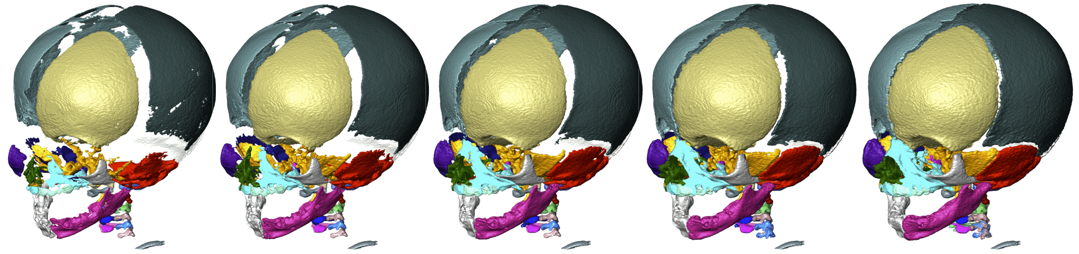
Computed tomography (CT) scanning is a standard way of visualizing hard tissues in living organisms. However, tridimensional reconstruction of CT images requires segmenting the structure at hand, which is time-consuming at best (i.e., manual segmentation) and imprecise at worst (i.e., automatic segmentation), especially in multipart segmentation. To circumvent this issue, Didziokas et al. (2024) have developed an open-access, user-friendly, automated segmentation tool for hard tissues, focusing especially on skull bones: boundary-preserving threshold iteration (BounTI). As its name suggests, BounTI’s operators select the structure of interest based on voxel intensity, which is the only input parameter it needs from the user. This procedure yields good results for bone segmentation, given that osseous tissue usually presents a distinct voxel intensity when compared with its surroundings. An appropriate initial threshold of voxel intensity is one that does not cause separate elements to be joint in the seed (that is, the first recognition of tissue by the algorithm), and which does not cause erroneous separations of single elements (e.g., the parietal bone) in the final stage.

BounTI was tested on skull CT images of various species, including amphibians, reptiles, and mammals. The quality of the assessment’s results demonstrates BounTI’s versatility and effectiveness. However, its performance is bound by the quality of the image; lower resolutions yield worse results. To mend any errors that might arise, BounTI does include options for manual intervention. Lastly, the authors emphasize the tool’s accessibility, human and machine-wise. BounTI can be implemented in a plethora of ways, holding great potential to improve efficiency and accuracy in anatomical studies and clinical applications involving hard tissue segmentation.

Tim Schuurman


Cortex morphology, function and socio-ecology

image (1)Evolutionary studies of the effects of socio-ecological factors on brain morphology and function, whether in extant or extinct species, have often led to contradictory results. In order to address this issue and properly study the patterns and sources of cerebral cortex evolution, Schwartz et al. (2023) have developed a common analytical frame of reference, filling the gaps among the analyses of humans and other extant mammals, as well as the fossil record. Namely, the authors established a joint geometric representation of the cortex morphology and function of ninety species of mammals, including commonly used experimental model organisms such as mice or macaques.

Using this common framework, ancestral cortical morphology independent of overall brain size and function were then modeled and assessed in relation to several social and ecological parameters (e.g., group size and habitat, respectively). Socio-ecological adaptations during evolution are reflected in the morphology and function of the cerebral cortex. Particularly, findings show how, for example, group size correlates with the expansion of the anterior cingulate cortex, an area of the prefrontal default mode network considered integral in understanding the intentions of others.

Nevertheless, despite Schwartz et al.’s (2023) incredible work, this research is not free from limitations; most notably, although inevitable, simplification. For instance, the large scope of the sample demands a reliance on single, often post-mortem, specimens of each of the studied species. This issue may have caused errors in their 3D modeling. Luckily, thanks to the increasing availability of open image datasets, this problem may be overcome in the future. The authors are aware of these limitations, and state their hopes for a refinement of their common reference frame of mammal cortical geometry in the future.

Tim Schuurman